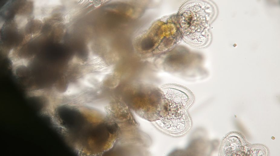
Cilia and flagella have a common structure consisting of an axoneme, or main cylinder of tubules, about 0.25µm in diameter. The axoneme is connected to a basal body and surrounded by an extension of the cell membrane. Between the axoneme and the basal body is a transition zone in which the arrangement of microtubules in the basal body takes on the pattern characteristic of the axoneme.
The basal body is identical in appearance to the centriole. A basal body consists of nine sets of tubular structures arranged around its circumference. Each set is called a triplet because it consists of three tubules that share common walls — one complete microtubule and two incomplete tubules. As a cilium or flagellum forms, a centriole migrates to the cell surface and makes contact with the plasma membrane. The centriole then acts as a nucleation site, initiating polymerisation of the nine outer doublets of the axoneme. After the process of tubule assembly has begun, the centriole is then referred to as a basal body.
The axoneme has a characteristic “9 + 2” pattern, with nine outer doublets of tubules and two additional microtubules in the centre, often called the central pair. The nine outer doublets of the axoneme are thought to be responsible for the sliding of adjacent doublets. The inter-doublet links (nexin connections) join adjacent doublets and the radial spokes project inward, terminating near projections that extend outward from the central pair of MTs. Each outer doublet of the axoneme therefore consists of one complete MT, called the A tubule, and one incomplete MT, the B tubule. The A tubule has 13 proto-filaments, whereas the B tubule has only 10 or 11. The tubules of the central pair are both complete, with 13 proto-filaments each. All of these structures contain tubulin, together with a second protein called tektin. Tektin is related to intermediate filament proteins and is a necessary component of the axoneme. The A and B tubules share a wall that appears to contain tektin as a major component.
Advertisement
In addition to microtubules, axonemes contain several other key structures. The most important of these are the sets of side arms that project out from each of the A tubules of the nine outer doublets. Each sidearm reaches out clockwise toward the B tubules of the adjacent doublet. These arms consist of axonemal dynein, which is responsible for sliding MTs within the axoneme past one another to bend the axoneme. The dynein arms occur in pairs, one inner arm and one outer arm, spaced along the MT at regular intervals. At less frequent intervals, adjacent doublets are joined by inter-doublet links. These links are thought to limit the extent to which doublets can move with respect to each other as the axoneme bends. At regular intervals, radial spokes project inward from each of the nine MT doublets, terminating near a set of projections that extend outward from the central pair of microtubules. These spokes are thought to be important in translating the sliding motion of adjacent doublets into the bending motion that characterises the beating of these appendages.
The writer is associate professor, head, department of botany, ananda mohan college, kolkata, and also fellow, botanical society of bengal, and can be contacted at tapanmaitra59@yahoo.co.in.
Advertisement


