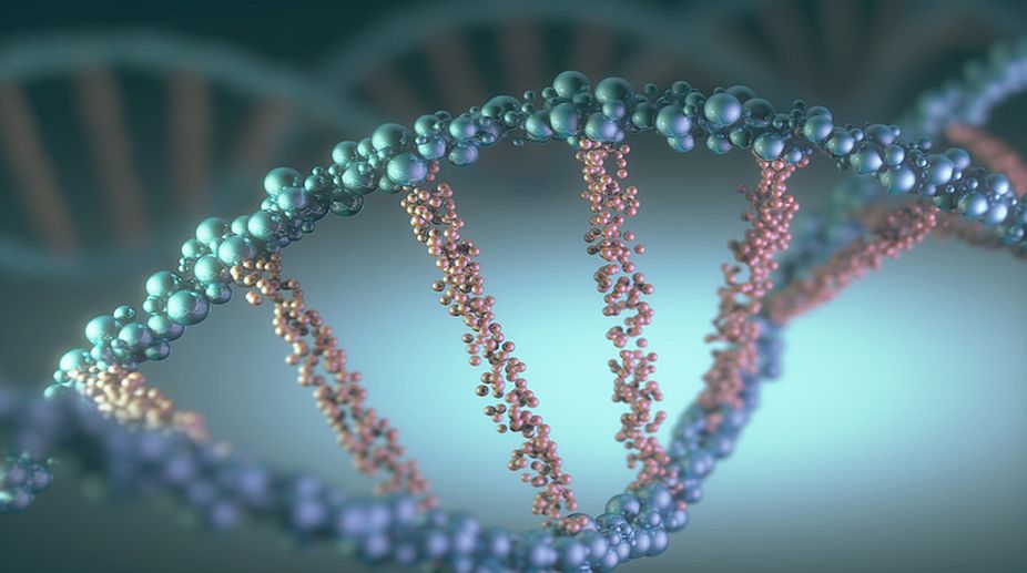
Maharashtra bus accident: Bodies to be identified through DNA testing
According to officials, the bus was travelling from Maharashtra's Yavatmal to Pune and met with an accident in Buldhana on the Samruddhi Mahamarg Expressway.

Representational Image (GETTY IMAGES)
It would obviously create problems if cells proceeded from one phase of the cycle to the next before the preceding phase had been properly completed. For example, if chromosomes start moving toward the spindle poles before they have all been properly attached to the spindle, the newly-forming daughter cells might receive extra copies of some chromosomes and no copies of others.
Similarly, it would be potentially hazardous for a cell to begin mitosis before all of its chromosomal DNA had been replicated. To minimise the possibility of such errors, cells utilise a series of checkpoint mechanisms that monitor conditions within the cell and transiently halt the cell cycle if conditions are not suitable for continuing.
The checkpoint pathway that prevents anaphase chromosome movements from beginning before the chromosomes are all attached to the spindle is called the spindle checkpoint. It works through a mechanism in which chromosomes whose kinetochores remain unattached to spindle microtubules produce a “wait” signal that inhibits the anaphase-promoting complex.
Advertisement
As long as the anaphase-promoting complex is inhibited, it cannot trigger destruction of the cohesins that hold sister chromatids together. The “wait” signal responsible for inhibiting the anaphasepromoting complex is transmitted by proteins that are members of the Mad and Bub families. The Mad and Bub proteins accumulate at unattached chromosomal kinetochores, where they are converted into a multi-protein complex that inhibits the anaphase-promoting chromosomes that have become attached to the spindle. This “wait” signal ceases and the anaphase-promoting complex becomes active.
A second checkpoint mechanism, called the DNA replication checkpoint, monitors the state of DNA replication to ensure that DNA synthesis is completed prior to permitting the cell to exit from G2 and begin mitosis. The existence of this checkpoint has been demonstrated by treating cells with inhibitors that prevent DNA replication from being finished. Under such conditions, the final de-phosphorylation step involved in the activation of mitotic Cdk-cyclin is blocked through a series of events triggered by proteins associated with replicating DNA. The resulting lack of mitotic Cdk-cyclin activity halts the cell cycle at the end of G2 until all DNA replication is completed.
A third type of checkpoint mechanism is involved in preventing cells with damaged DNA from proceeding through the cell cycle unless the DNA damage is first repaired. In this case, a multiple series of DNA damage checkpoints exist that monitor for DNA damage and halt the cell cycle at various points — including late Gl, S, and late G2 — by inhibiting different Cdk-cyclin complexes. A protein called p53, sometimes referred to as the “guardian of the genome,” plays a central role in these checkpoint pathways.
When cells encounter agents that cause extensive DNA damage, the altered DNA triggers the activation of an enzyme called ATM protein kinase, which in turn catalyses the phosphorylation of p53 (and several other target proteins). Phosphorylation of p5i prevents it from interacting with Mdm2, a protein that would otherwise mark p53 for destruction by linking it to ubiquitin (just as the anaphase-promoting complex target proteins for degradation by linking them to ubiquitin) ATM-catalysed phosphorylation of p53 therefore protect it from degradation and leads to a buildup of p53 in the presence of damaged DNA.
The accumulating p53 in turn activates two types of events — cell-cycle arrest and cell death. Both responses are based on the ability of p53 to bind to DNA and act as a transcription factor that stimulates the transcription of specific genes. But if the damage cannot be successfully repaired, p53 then activates a group of genes coding for proteins involved in triggering cell death by apoptosis. A key protein in this pathway, called Puma (“p53 unregulated modulator of apoptosis”), promotes apoptosis by binding to and inactivating a normally occurring inhibitor of apoptosis known as Bcl-2.
The writer is associate professor, head, department of botany, ananda mohan college, kolkata, and also fellow, botanical society of bengal, and can be contacted at tapanmaitra59@yahoo.co.in.
Advertisement