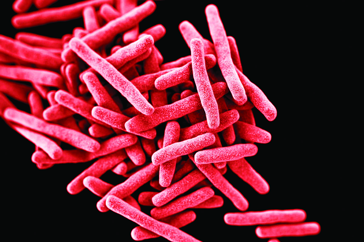
The organism responsible for leprosy, Mycobacterium leprae, was discovered in 1874 by the Norwegian investigator Gerhard Hansen. Leprosy, however, was well known even in Ancient Egypt. In the Middle Ages and during the Crusades, it spread as an epidemic, since that period was characterised by continuous wars that caused bad sanitary conditions. There were 2,000 leper colonies in France in 1429 but the disease disappeared from Europe at the end of the 17th century. In France, all leper colonies were closed on 24 August 1693. An increase in disease incidence occurred again from 1867, followed by a marked decline at the beginning of the 20th century. Disease prevalence, however, is still high in these times.
Infection source and route
The source of infection is a sick person and the causative agent is transmitted by the air droplet route through the nasopharynx and injured skin. The infection may also be spread by various objects. Intimate and prolonged contact between healthy individuals and leprosy patients is, however, the main mode of infection.
After entering the body through the skin and mucous membranes, the organisms penetrate the nerve endings, lymphatic and blood vessels, and disseminate gradually without causing any changes at the site of entry. In the presence of high body resistance, the majority of M. leprae perish. In some cases, infection leads to the development of latent forms of leprosy.
The duration of such latent forms depends on body resistance, and may persist for a lifetime and, as a rule, terminates with the death of the causative agent. The latent form may change to the active form with development of the disease, if living and working conditions become unfavourable. The incubation period may last for years, may be from a period of three to five or 20-35 years.
Advertisement
Types of leprosy
Three types of leprosy are distinguished based on clinical manifestations — lepromatous, tuberculoid and undifferentiated. The lepromatous type is characterised by minimum body resistance to the presence, multiplication and spread of the causative agent. M. leprae are constantly present at the sites of the lesions and the lepromin skin test is negative.
On the other hand, the tuberculoid type is distinguished by high body resistance to the multiplication and spread of M. leprae. Either no organism is found at the site of lesions, or only a small number of them may be present during the reactive state. The allergic test is usually positive.
Finally, the undifferentiated type (non-specific group) is characterised by varying body resistance. Microscopic examination does not always reveal the presence of M. leprue and allergic tests are negative or yield a slightly positive reaction.
Immunity
Little is known about immunity in connection with leprosy, but an allergic condition develops during the disease. The mechanism of immunity in leprosy is like that in tuberculosis.
In individuals with high body resistance, organisms are phagocytosed by histiocytes in which they are destroyed quite rapidly. In such cases leprosy assumes a benign tuberculoid type. In individuals with low resistance, M. leprae multiply in great numbers even within the phagocytes (incomplete phagocytosis), and the organisms disseminate throughout the body. A severe lepromatous type of the disease develops in such individuals.
Resistance may vary from high to low in undifferentiated types of leprosy. Relatively benign lesions persist for years, but if body resistance lowers, the disease assumes a lepromatous form with numbers of mycobacteria present in the tissues and organs. The clinical picture changes to the tuberculoid type when immunity intensifies.
Immunity in leprosy is associated with the general condition of the host body. In most cases, the disease occurs among people living in unhygienic conditions and children are most susceptible. In five per cent cases, the disease is acquired through contact with sick parents.
Laboratory diagnosis
Specimens for examination are obtained from nasal mucosa scrapings (on both sides), skin lepromas, sputum and ulcer excretions. Blood is examined during the fever period and microscopic examination is the principal method of leprosy diagnosis. The biopsy of leprotic lesions and puncture of lymph nodes are employed in some cases. M. leprae can be seen as clusters resembling packets of cigars; in preparations from nasal mucus they appear as red balls. The allergic Mitsuda test is considered positive when an erythema and a small papule (early reaction) are produced at the site of an 0.1 millimetre lepromin (a suspension prepared from a leproma after trituration and prolonged boiling) injection in 48 to 72 hours. This reaction either disappears completely at the end of the first week or changes to the late reaction. The latter is manifested by a nodule, which appears at the site of injection in 10-14 days and grows to a diameter of one to two cm with necrosis at the centre. This test, however, is of no diagnostic value and is used to distinguish the clinical type of leprosy. The complement-fixation reaction and the Middlebrook-Dubos Haemagglutination test are employed for leprosy diagnosis.
Treatment
Leprosy is treated with sulphone drugs (dapson), and diaminodiphenylsulphone and its derivatives (sulphetrone, promin, diazone and promacetin). In addition to that, conteben, desensitising agents, and corticosteroid preparations (cortisone, prednisolone, etc) are employed.
For a long period of time, leprosy patients were treated with chaulmoogra oil. At present it is administered intramuscularly or intracutaneously. Chaulmoogra preparations promote the resolution of lesions and, sometimes, eliminate visible leprosy manifestations. They, however, give no protection from relapses and have no specific effect.
According to the World Health Organisation, more than 10 million people suffering from leprosy are registered throughout the world — 6.4 million in Asia, 3.8 million in Africa, 385,000 in America, 52,000 in Europe and 33,000 in Oceania. The high prevalence of leprosy makes research into the methods of its specific prophylaxis necessary.