New research claims Mediterranean diet helps boosts IVF success rate
For more than 20 years, millions of women have successfully utilized In vitro fertilization (IVF) as a reproductive treatment.
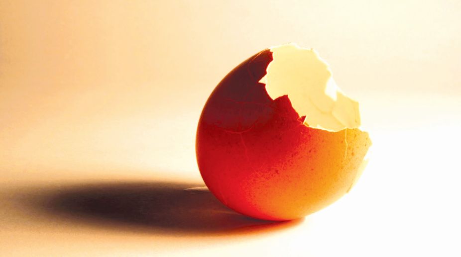
Reptile and bird eggshells take good care of the embryo but let go when it’s time to hatch.

Eggshell
The eggshell’s first function is protection while the fertilised egg develops. During this time, the eggshell also dissolves from the inside, to be a source of calcium for the growing embryo. This thinning of the inner layers of the shell structure, in turn, helps weaken the shell, so that it cracks when the chick is ready to emerge.
Dimitra Athanasiadou, Wenge Jiang, Dina Goldbaum, Aroba Saleem, Kaustuv Basu, Michael S Pacella, Corinna F Böhm, Richard R Chromik, Maxwell T Hincke, Alejandro B Rodríguez-Navarro, Hojatollah Vali, Stephan E Wolf, Jeffrey J Gray, Khanh Huy Bui and Marc D McKee, from McGill University, Montreal; John Hopkins University, Baltimore; Friedrich-Alexander-University, Germany, and the University of Ottawa, describe in the journal, Science Advances, their study of how the nano-structure of the eggshell forms and how it facilitates its diverse functions during the incubation period.
Advertisement
And then, the team shows that the protein that brings about the eggshell structure can create structure in the non-biological material, just like it does in the eggshell.
Advertisement
Reptile and bird eggshells are remarkable containers that allow the embryo to develop in the rich nutrients of the egg, and in safety. The researchers discover that the eggshell is not the same at all parts of its thickness, and it is a protein called osteopontin, which was first found to help bone tissue to form, that gives the eggshell its structure.
Osteopontin is a chain molecule that has many points along its length where it shows a negative charge. Atoms of calcium and minerals bind to these points along the protein molecule. Osteopontin thus forms a scaffold and the calcium carbonate (the same thing as chalk), which forms 95 per cent of the eggshell, grows into a matrix, to form a shell with the optimum structure that makes for strength.
“Eggshells are notoriously difficult to study by traditional means, because they easily break when we try to make a thin slice for imaging by electron microscopy,” a press release reports McKee from McGill University to have said. “Thanks to a new focused ion beam sectioning system recently obtained by McGill’s Facility for Electron Microscopy Research, we were able to accurately and thinly cut the sample and image the interior of the shell.”
Using these methods, the team finds that the eggshell material of the eggs of the domestic chicken (Galus gallus) consists of nano-granules, whose size varies as one passes from the outer to the inner surface.
The paper in Science Advances explains that living things create a variety of biominerals, in the form of hardened structures, like human bones. In human bones, the material is mainly nano-crystals of calcium phosphate, which forms within a matrix of large organic molecules. In the case of other organisms, the material is mainly calcium carbonate (limestone or chalk), which form rigid structures, typically shells.
In the case of shells of mollusk, coral skeletons, the granular structure has generally been found to be uniform, with the material behaving as if it were just one crystal. In the case of the eggshell, analysis has revealed a varying pattern in the grain size as one passes though the thickness of the shell.
It is found that in the outermost layer, called the vertical crystal layer, the grain size is 30 nanometres. The upper palisades layer, where grain size is 33 nm, comes next. The grain size rises again to 59 nm in the middle PL, to 74 nm in the lower PL and 68 nm in the innermost mammillary layer. This variation in the granular size was found to correspond to the levels of osteopontin present — a large presence in the VCL resulted in fine grain size.
While the gradient in grain size was noted, further study of the PL region, the largest in the section of the eggshell, revealed a pattern of clustering of domains from five to seven nm in size.
As a mechanism of how the pattern forms, the researchers suggest that it is similar to the formation of patterns in distribution of pigment, as in leopards’ spots or tigers’ stripes, and also related to the abundance of osteopontin during different stages of the eggshell formation.
The team also studied the progression of hardness and elasticity of the eggshell material. It was found that the outermost VCL layer was the hardest and most elastic, with the properties reducing till the middle of the PL, and then rising towards the innermost of the ML.
That the hardness was highest in the VCL, where the grain size was the smallest is in keeping with the known principles of composite materials, but the rise in hardness towards the ML is still not understood, the study says.
The first understanding from the study is the structure of the avian eggshell and its twin roles — the inner layer being easily dissolved to provide the embryo with calcium for bone development and thereby weakening so that the chicken can hatch when it is ready.
But an important finding is that the shell strength can be varied by quantity of osteopontin that is present. It was shown by the team that even outside a biological setting, the mineralising of calcium carbonate was just like in the eggshell when osteopontin was present. And then, more osteopontin led to finer grain size, as in the eggshell.
The paper says that a problem that the poultry industry faces is that when a hen has been laying eggs for about a year, the shells start getting brittle. This could allow salmonella infection and food safety. Knowledge about the eggshell structure and the role of osteopontin could suggest genetic engineering methods for stronger eggshells.
Understanding the role of organic components in structure of composite materials would also help “inform design concepts for synthetic nanocomposites that have novel properties,” the study says.
The writer can be contacted at response@simplescience.in
Advertisement