Elon Musk shares health advice on severe neck and back pain
Disc replacement surgery may be a game-changer for people experiencing severe neck or back pain, said billionaire Elon Musk on Tuesday.
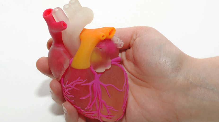
Cardiologists preparing to perform life-saving heart valve replacements can now use customisable 3D-printed models of the organ to assist them…
(Photo: Getty Images)
Cardiologists preparing to perform life-saving heart valve replacements can now use customisable 3D-printed models of the organ to assist them with the surgeries, scientists say.
Researchers at Georgia Institute of Technology and the Piedmont Heart Institute in the US are using standard medical imaging and new 3D printing technologies to create patient- specific heart valve models that mimic the physiological qualities of the real valves.
The aim is to improve the success rate of transcatheter aortic valve replacements (TAVR) by picking the right prosthetic and avoiding a common complication known as paravalvular leakage.
Advertisement
“Paravalvular leakage is an extremely important indicator in how well the patient will do long term with their new valve,” said Zhen Qian, from Piedmont Heart Institute.
“The idea was, now that we can make a patient-specific model with this tissue-mimicking 3D printing technology, we can test how the prosthetic valves interact with the 3D printed models to learn whether we can predict leakage,” Qian said.
The study, published in the journal JACC: Cardiovascular Imaging, found that the models created from CT scans of the patients' hearts behaved so similarly to the real ones that they could reliably predict the leakage.
“These 3D printed valves have the potential to make a huge impact on patient care going forward,” said Chuck Zhang, a professor at Georgia Tech.
“Our 3D printed model gives us a quantitative method to evaluate how well a prosthetic valve fits the patient,” Qian said.
The models are created with a special metamaterial design and then made by a multi-material 3D printer, which gives the researchers control over such design parameters as diameter and curving wavelength of the metamaterial used for printing, to more closely mimic physiological properties of the tissue.
“Previous methods of using 3D printers and a single material to create human organ models were limited to the physiological properties of the material used,” Zhang said.
“Our method of creating these models using metamaterial design and multi-material 3D printing takes into account the mechanical behaviour of the heart valves, mimicking the natural strain-stiffening behavior of soft tissues that comes from the interaction between elastin and collagen, two proteins found in heart valves,” he said.
“Eventually, once a patient has a CT scan, we could create a model, try different kinds of valves in there, and tell the physician which one might work best,” Qian said.
“We could even predict that a patient would probably have moderate paravalvular leakage, but a balloon dilatation will solve it,” he said.
Advertisement